


Medical Imaging: an Essential Component of Real-World Data
Reading time /
4 min
Industry
.png)
TL;DR
- Medical imaging provides ground-truth visual evidence of anatomy and physiology that written records cannot replicate making it a foundational component of real-world data.
- Longitudinal imaging data allows researchers to track disease progression and treatment response over time, across the full arc of a disease course or treatment program.
- Segmed’s network covers the full spectrum of diagnostic imaging modalities, all de-identified to HIPAA Safe Harbor standards and delivered in machine-ready DICOM format.
- Real-world imaging data supports AI model training, clinical trial feasibility, RWE generation, drug development, and FDA and CE regulatory submissions.
Introduction
Medical imaging is a core component of modern clinical practice and an increasingly critical input for healthcare research. From X-rays and CT scans to MRI, PET, and digital pathology, imaging provides visual evidence of anatomy and physiology that written records cannot replicate. Segmed aggregates this imaging evidence at scale, sourced from healthcare partner sites across the U.S. and five continents, and makes it accessible for research, AI development, and regulatory submissions.
In recent years, there has been a growing focus on using real-world data (RWD) and real-world evidence (RWE) in healthcare and medical research. Real-world data comes from a variety of sources, including electronic health records, insurance claims data, patient registries, and medical imaging data. The sections below describe why imaging data occupies a foundational role in real-world evidence generation.
The Power of Imaging Ground Truth
In domains such as radiology, dermatology, ophthalmology, and pathology, the image is a reference ground-truth data. Medical imaging provides visual evidence that can be far more persuasive and insightful than clinical descriptions alone, especially in contexts where two medical experts often disagree. Seeing a tumor, organ abnormality, or site of injury gives physicians a clearer understanding of patient conditions and helps them agree on a diagnosis. Access to patient imaging data, versus just reports or summaries, allows researchers to make direct observations that can lead to discoveries.
Connecting Symptoms to Structures
Patients often present with ambiguous symptoms like pain, fatigue, dizziness, that many different underlying conditions could cause. Medical imaging allows physicians to investigate the anatomical source of those symptoms. For example, persistent headaches may be explained by MRIs that reveal a brain tumor. With imaging data, physicians can match symptoms to physical structures and pathologies to accurate diagnoses and treatment plans.
Tracking Changes Over Time
Many diseases manifest through subtle changes in the body over weeks, months, or years. Medical imaging allows physicians to visually track these changes by comparing scans taken at different time points. Multiple CT chest scans, for example, can show the progression of lung cancer tumors and quantify changes in size. Access to longitudinal imaging gives researchers crucial insight into how a patient’s condition evolves, across the arc of a disease course or treatment program.
Imaging as Regulatory Evidence
Medical imaging data from real-world sources can provide additional clinical trial endpoints beyond typical measures such as survival rates or laboratory results. Imaging can visually demonstrate the effects of investigational drugs and devices. Access to imaging data at all stages of research from early phases through post-market studies can further confirm product safety and efficacy. The FDA recognizes medical imaging as a biomarker supporting regulatory decision-making, and imaging data sourced from real-world clinical populations is increasingly used to support IND, NDA, and PMA submissions.
Research Use Cases by Application and Customer Type
The value of real-world imaging data varies by research context. Below are the primary applications Segmed’s imaging network supports:
Understanding Total Patient Health
When analyzing real-world data, it is essential to look at the complete health profile of patients. Medical records alone may not provide sufficient information to paint a full picture. Imaging data fills in gaps by illuminating patient anatomy and physiology directly. Radiologists analyze images for incidental findings that may be meaningful to patient health, even if unrelated to the original imaging indication. Access to complete imaging data ensures research provides a holistic view of patient health trajectories.
Big Data Drives Big Discoveries
The value of real-world evidence lies in aggregating and analyzing large volumes of data from diverse sources. Every medical image represents an extensive set of data points that can be applied to population health studies using large-scale analysis techniques. Imaging datasets enhanced by AI are producing documented findings about disease progression, treatment effects, and precision diagnostics. Harnessing large-scale imaging data is a prerequisite for maximizing the utility of real-world evidence in both clinical research and product development.
Imaging Modalities and Associated Data Types Available for Research
Effective real-world imaging research requires data that reflects the modality diversity present in clinical practice. Segmed’s network covers the full spectrum of diagnostic imaging modalities in active clinical use:
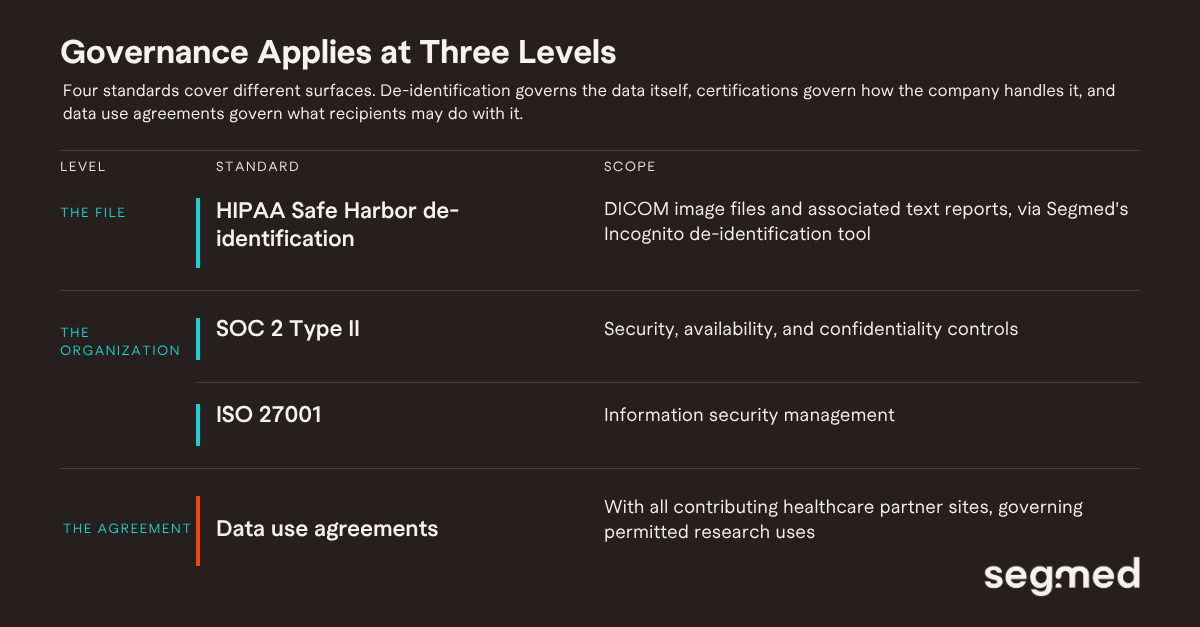
Each imaging study in the Segmed network is paired with available associated data including radiology reports, EHR-derived diagnoses, treatment history, outcomes, and patient demographics enabling multimodal analysis across imaging and clinical variables. All data is delivered in DICOM format, de-identified to HIPAA Safe Harbor standards, and structured for machine readiness.
Compliance, De-identification, and Data Governance
Access to real-world imaging data for research requires that privacy and compliance requirements are met before data reaches the researcher. Segmed handles de-identification and governance end-to-end, so research teams receive data that is analysis-ready and audit-ready from day one.
Core compliance infrastructure includes:
For teams preparing regulatory submissions, including FDA and CE mark filings, Segmed’s datasets are structured to support the documentation requirements of regulatory-grade evidence packages.
Segmed’s Imaging Data Network
Medical imaging linked to clinical variables diagnoses, treatment history, outcomes, and demographics is the foundation of credible real-world evidence. Segmed customers including Bayer, GE Healthcare, Microsoft, Johnson & Johnson, Siemens, and Harvard access this data through Openda, Segmed’s self-service imaging data platform, which provides structured search across a large-scale imaging network and supports cohort export in machine-ready formats.
Ready to explore fit-for-purpose RWiD for your program? Connect with us to discuss your data requirements or submit a project feasibility inquiry to evaluate modality and cohort availability for a specific program.
Frequently Asked Questions - F.A.Q
What is real-world imaging data?
Real-world imaging data is medical imaging CT scans, MRI, X-rays, PET, digital pathology, and other modalities collected during routine clinical care rather than in controlled trial settings. It reflects the full diversity of clinical practice: different scanner types, acquisition protocols, patient populations, and care settings. This diversity is what makes it valuable for research that needs to generalize beyond a single institution.
How is medical imaging different from other types of real-world data?
Unlike electronic health records or claims data, medical imaging provides direct visual evidence of anatomy, pathology, and physiology. Images capture information that cannot be adequately described in written records tumor morphology, organ structure, lesion location and size. In clinical domains such as radiology, dermatology, and pathology, the image is the ground truth.
What imaging modalities does Segmed’s network cover?
Segmed’s network covers the full spectrum of diagnostic imaging in active clinical use: CT, MRI, X-ray, PET/CT, PET/MR, ultrasound, fluoroscopy, digital pathology with whole-slide imaging, mammography and breast imaging, and ophthalmology imaging. All studies are delivered in DICOM format, de-identified to HIPAA Safe Harbor standards.
How does Segmed ensure compliance and de-identification?
Segmed applies HIPAA Safe Harbor de-identification to both DICOM image files and associated text reports via its Incognito tool. The platform operates under SOC 2 Type II and ISO 27001 certifications. Data use agreements govern all contributing healthcare partner sites. Research teams receive data that is analysis-ready and audit-ready from day one.
What research use cases does real-world imaging data support?
Real-world imaging data supports AI model training and validation, clinical trial feasibility and endpoint definition, post-market surveillance, comparative effectiveness research, drug development and radiomics, and academic or CRO research across rare diseases and underrepresented populations. It can also support regulatory submissions including FDA 510(k), De Novo, IND, NDA, and PMA pathways.
Which companies use Segmed’s imaging data?
Segmed customers include Bayer, GE, Microsoft, Johnson & Johnson, Siemens, and Harvard, among others. They access imaging data through Openda, Segmed’s self-service imaging data platform, which provides structured search and supports cohort export in machine-ready formats.
How is imaging data used to support regulatory submissions?
The FDA recognizes medical imaging as a biomarker supporting regulatory decision-making. Real-world imaging data sourced from clinical populations is used to support IND, NDA, and PMA submissions, as well as FDA 510(k) and De Novo filings for AI-enabled medical devices. Segmed datasets are structured with the provenance and documentation requirements of regulatory-grade evidence packages.
References
Fit-for-purpose real-world imaging data: the difference between more data and better data
White Paper: A Framework for fit-for-purpose real-world imaging data
Multimodal data pipelines: the new gold standard in pharma research